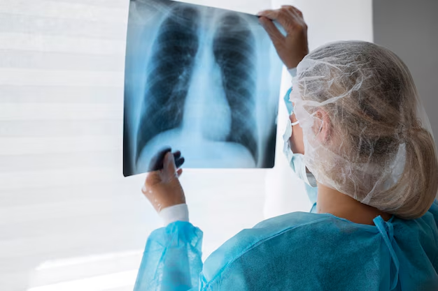
Lung Cancer
Lung cancer is one of the leading causes of cancer-related deaths globally. It can develop in any part of the lungs and is often linked to smoking, pollution, and genetic factors. Early detection is critical for successful treatment. Symptoms may include a persistent cough, chest pain, unexplained weight loss, or coughing up blood. Dr. Manoj Kumar Goel, an experienced pulmonologist and interventional specialist in Delhi, offers advanced diagnostic tools such as bronchoscopy, EBUS, and high-resolution imaging along with a multidisciplinary approach for effective lung cancer care and management.
Diagnosing Lung Cancer with Bronchoscopy and Lung Biopsy
Bronchoscopy with lung biopsy is a key procedure for diagnosing lung cancer. Bronchoscopy involves placing a thin tube-like instrument called a bronchoscope through the nose or mouth and down into the airways of the lungs. The tube has a mini-camera at its tip, which carries pictures back to a video screen or camera. Using the bronchoscope, the doctor can see the interior of the airway, identify any abnormalities, and decide on the type of sample to be taken. Depending on the findings, a biopsy forceps or a cryo probe is introduced via the working channel of the bronchoscope, and a Transbronchial Lung Biopsy (TBLB), cryobiopsy, or endobronchial biopsy is performed. In selected cases, the biopsy may be done under the guidance of a radial probe EBUS for improved accuracy.
Possible Risks and Complications of Bronchoscopy with Lung Biopsy
Every effort will be made to conduct the procedure in such a way as to minimize discomfort and risks. However, as with other types of procedures, there are inherent potential risks.
-
General Complications: The incidence of major complications associated with bronchoscopy is 0.8% - 1.3%. These include accumulation of air in the pleural space, hemorrhage, subcutaneous emphysema, postoperative fever, chest infection, cardiac arrhythmias, hypoxemia, vasovagal attack, myocardial infarction, pulmonary edema, bronchospasm, choking, and perforation of the airway.
-
Cryobiopsy-Specific Risk: The risk of pneumothorax with cryobiopsy is 5%.
-
Mortality Rate: The mortality rate associated with bronchoscopy is less than 0.01%.
Post-Procedure Course for Bronchoscopy with Lung Biopsy
-
Postoperative oxygen supplementation may be required in some patients, particularly those with impaired lung function and those who have been sedated.
-
A chest radiograph is carried out post-procedure to monitor for complications.
-
Patients who have had transbronchial biopsies should observe for pain chest, breathlessness, haemoptysis, surgical emphysema, or excessive cough, which can indicate pneumothorax after leaving the hospital, and they should contact hospital emergency.
-
Patients who have been sedated are advised not to drive, sign legally binding documents, or operate machinery for 24 hours after the procedure.
-
It is preferable that day case patients who have been sedated should be accompanied home, and higher-risk patients, such as the elderly or those from whom transbronchial biopsy specimens have been taken, should have someone to stay with them at home overnight.


